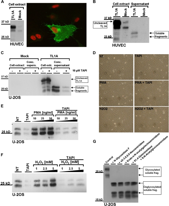
Figure 2.
Ectodomain shedding of transmembrane tumor necrosis factor–like cytokine 1A (TL1A) in human umbilical vein endothelial cells (HUVEC) and U-2OS cells. (A) HUVEC were transfected with TL1A cDNA or empty vector (Mock) by AMAXA electroporation, immunostained for TL1A, and examined by confocal fluorescence microscopy. Nuclei were stained with TO-PRO 3 (right). Expression of TL1A was verified by Western blot (left). (B) HUVEC were transfected with either empty vector (Mock) or a TL1A overexpression vector by AMAXA electroporation. HUVEC cell extracts and supernatant were examined by Western blot using antibodies specific for TL1A. (C) U-2OS cells were transiently transfected with TL1A cDNA and Mock using Lipofectamine 2000. After antibiotic selection for transfected cells, they were seeded at a density of 3 × 10⁵ per well in a six-well plate. Ten micromolars of TAPI-1 and the equivalent volume of DMSO as a control were added as indicated and incubated for 24 hours. Cell extracts and speed-vac concentrated supernatants were examined by Western blot using anti-TL1A antibody. (D) U-2OS cells were treated for 2 hours with 50 ng/mL phorbol-12-myristat-13-acetate (PMA) and 5 mM H2O2, as indicated. Micrographs of treated cells are shown. (E and F) U-2OS cells were transfected with TL1A cDNA and seeded as described previously. Cells were either untreated (NT) or treated with increasing concentrations of PMA (E) or H2O2 (F) in the presence or absence of TAPI-1 (20 μM), as indicated. After 2 hours, supernatants were harvested, concentrated by speed-vac evaporation, and subjected to Western blot analysis. (G) Supernatant of U-2OS cells overexpressing TL1A was concentrated via speed-vac evaporation and treated with the indicated enzymes to remove all N- and O-linked oligosaccharides. The enzymes were added sequentially, in a cumulative manner, so that in the end all five enzymes are added to the reaction (from left to right on the indicated Western blot). TL1A fragments were separated on a large sodium dodecyl sulfate–polyacrylamide gel electrophoresis for higher resolution and analyzed by Western blot using anti-TL1A antibody.

