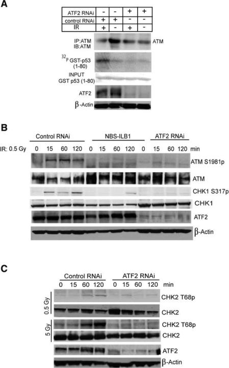
Figure 6. ATF2 Is Important for IR-Induced Activation of ATM and Chk2.
(A) ATF2 is important for activation of ATM. U2OS cells were infected with either control or ATF2 RNAi, and 72 hr later, cells were subjected to IR (6 Gy). Proteins prepared 1 hr after IR were subjected to IP with antibodies to ATM. Immunoprecipitated material was used for immunokinase reactions using GST-p531–80 as a substrate. Upper panel depicts the autoradiograph, whereas lower panels show input and level of ATF2 as well as β-actin expression.
(B) ATF2 and Nbs1 are important for ATM and Chk1 activation by IR. GM00637 cells were subjected to infection with control or ATF2 RNAi and were analyzed in parallel to NBL-ILB1 cells at the indicated time points following a low dose of IR (0.5 Gy) for ATM activation using phosphorylation of ATM on Ser1981 (upper panel) and the phosphorylation of Chk1 on Ser317 (third panel). Also shown are total ATM (second panel), total Chk1 (fourth panel), ATF2 expression (fifth panel), and β-actin, used as a loading control (lower panel).
(C) Inhibition of ATF2 expression impairs IR-induced Chk2 phosphorylation. Control or ATF2 RNAi-infected cells were treated by IR (0.5 Gy or 5 Gy) 72 hr postinfection and proteins prepared at the indicated time points. Immunoblot analysis using CHK2 antibodies detects phosphorylated (Thr 68; upper panel) and nonphosphorylated (second panel) forms of Chk2. Controls for the expression level of endogenous ATF2 and β-actin are shown.

