
FIGURE 1. ADAR double-stranded RNA binding domains and the KKXXK motif.
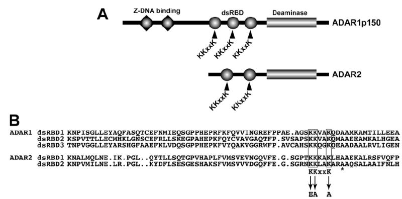
A, domain structure of ADAR1 and ADAR2 indicating Z-DNA binding domains (triangles), dsRBD (circles), and the C-terminal deaminase domain (rectangle). The KKXXK motif in each dsRBD is indicated. B, sequence alignment of the ADAR1 (top) and ADAR2 (bottom) dsRBD displaying the homology around the KKXXK motif and the mutated lysines to EAXXA (termed EAA). The asterisk denotes an alanine mutated in other studies.