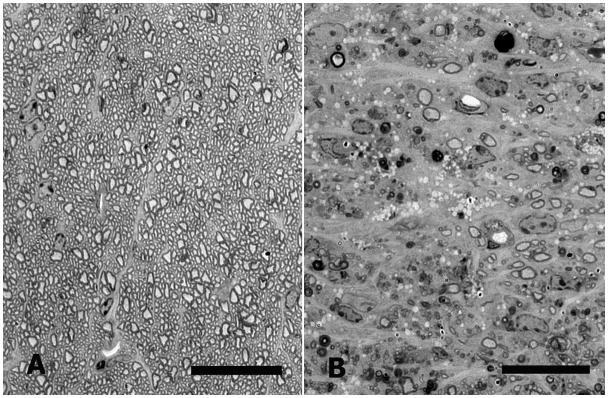
Figure 4.
Optic nerve cross-sections from CD1 mice. A: Normal mouse optic nerve. B: Nerve from bead-injected eye 12 weeks after injection, showing major loss of normal axon profiles, as well as clumps of myelin debris and macrophages filled with clear vacuoles (epoxy-embedded, 1 μm section, 1% toluidine blue, scale bars = 20 μm).

