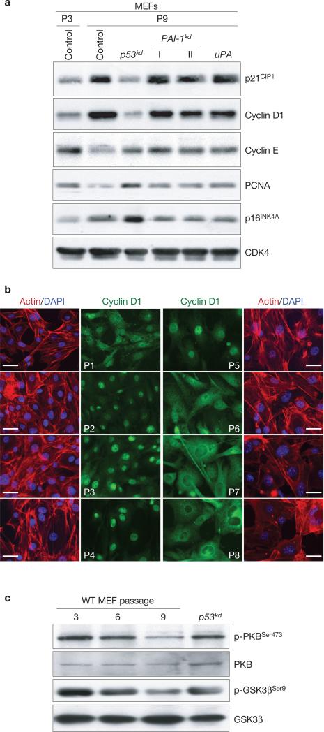
Figure 2.
MEFs reduce PKB activation and exclude cyclin D1 from the nucleus during replicative senescence. (a) Western blot analysis of P3, P9, p53kd, PAI-1kd or uPA overexpressing MEFs for cell-cycle related proteins. PCNA and CDK4 are proliferation and loading controls, respectively. (b) Qualitative immunofluorescence microscopy analysis of serially passaged MEFs, (P1–P8), for cyclin D1 expression. The scale bar represents 50 μm. (c) Expression of phosphorylated PKB or GSK3β related to unphosphorylated fraction of the same proteins in P3, P6 and P9 MEFs and post-senescent p53kd cells as analysed by western blot.

