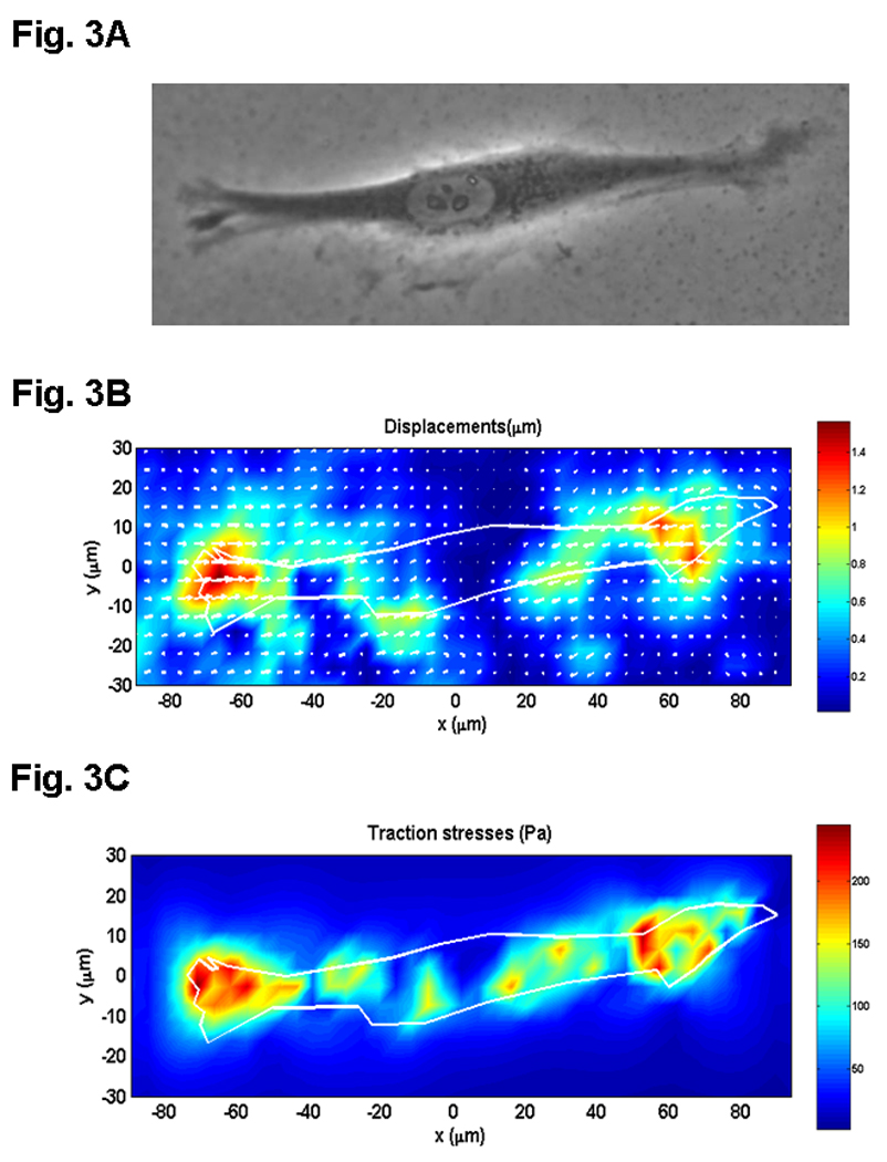
Fig. 3.

CTFM application to determine traction forces of human patellar tendon fibroblast (HPTF). A. A HPTF on PG substrate embedded with fluorescent microbeads, which are not shown on this phase contrast image. B. PG substrate displacements defined by beads’ movements near the PG surface. C. CTF field. (adopted with permission from Fig. 6 in Yang et al. 2006, J Theor Biol 242:607–616).
