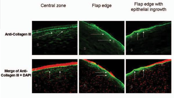
Figure 3.
Low expression of collagen III (arrows) in ectatic human corneas after LASIK surgery is observed at the central zone (left panels) and flap edge (middle panels) of corneas with subtle flap edges. Significant expression of collagen III (arrows) was observed in the peripheral fibrotic scar region (right panels) in the cornea with epithelial ingrowth. 4'6-diamidino-2-phenylindole (DAPI) was used for nuclear counterstaining (red cells). E=epithelium, S=stroma

