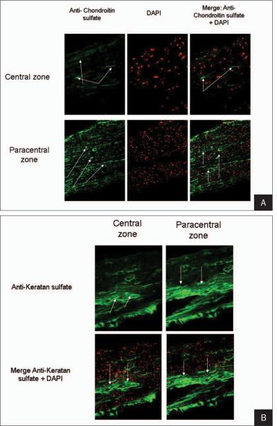
Figure 5.
A) Increased expression of chondroitin sulfate (arrows) is observed in the central zone (upper panels) compared with the paracentral corneal flap edge (lower panels). Left panels show the expression of chondroitin sulfate. Middle panels demonstrate 4'6-diamidino-2-phenylindole (DAPI) nuclear counterstaining (red cells). Right panels show a merge of both stains. B) Increased expression of keratan sulfate (arrows) is observed in the central zone (left panels) compared with the paracentral corneal flap edge (right panels). Upper panels show the expression of keratan sulfate. Lower panels illustrate a merge of keratan sulfate and 4'6-diamidino-2-phenylindole (DAPI) nuclear counterstaining (red cells).

