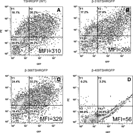
Figure 2.
Expression of TSHR subunits in HEK-293 cells. Expression and cellular localization for the full-length WT and the three truncated β-subunits was assessed by flow cytometry Cell surface expression of receptors was detected on unfixed cells with antibody M1 (residues 381-385) followed by staining with secondary antimouse PE (1:200). Due to the presence of GFP and PE, β-316 (panel B) showed 27% and β-366 showed 32% (panel C) of double-positive cells for surface expression that was similar to full-length TSHR (panel A). However, β-409 (panel D) showed a 29% GFP expression only, indicating just intracellular expression of the product. The FL2 mean fluorescence intensity (MFI) for each of the constructs is also indicated.

