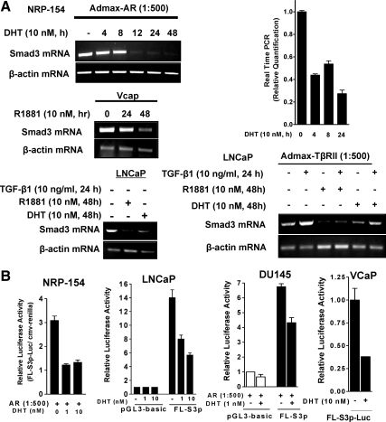
Figure 3.
Transcriptional regulation of Smad3 by DHT in NRP-154, LNCaP, DU145, and VCaP cells. A, Androgens repress Smad3 mRNA levels in prostate cancer cell lines. NRP-154 cells were plated in 100-mm dishes with modified GM3 medium containing 1% DC-FBS. After overnight for attachment, cells were and infected with AdMax-AR (1:500) for 2 h and then treated with DHT (10 nm, 4–48 h). VCaP cells were incubated with 10 nm of R1881 for 24 or 48 h. For LNCaP cells, the effect of DHT or R1881 (10 nm, 48 h) on Smad3 mRNA levels was examined either in noninfected cells, or in cells infected with AdMax-TβRII (1:500, 24 h), followed by TGF-β treatment for an additional 24 h. cDNA was made from 5 μg of total RNA (purified by RNeasy) by RT Superscript and semiquantitative PCR Smad3 expression (for NRP-154, LNCaP, and VCaP), or real-time PCR using TaqMan Real Time PCR kit and 7500-Real Time PCR System (Applied Biosystems) (for NRP-154 cells). B, Androgens repress activity of the Smad3 promoter in prostate cancer cell lines. Cells were transfected with a total 1 μg of DNA including either control luciferase vector (pGL3-basic) or full-length (1892 bp) Smad3 promoter-luciferase (FL-S3p-luc) construct and CMV-Renilla, followed by DHT treatment for 48 h. For NRP-154 and DU145 cells, cells were transfected in the presence of AR (AdMax-AR, 1:500). Data shown are relative values of firefly luciferase normalized to Renilla luciferase. Each bar represents the average of triplicate determinations ± se.

