Figure 4.
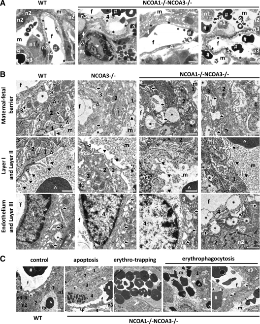
Abnormal morphogenesis observed in the labyrinths of E12.5 DKO mouse embryos. A, Toluidine blue-stained semithin sections of placental labyrinths from E12.5 embryos with the indicated genotypes. B, Subcellular ultrastructures of placental labyrinths from E12.5 embryos with the indicated genotypes. C, Several abnormalities observed in DKO labyrinths at E12.5. The image of a WT sample served as a control. Image annotations: 1, layer I mononuclear trophoblast; 2, layer II syncytiotrophoblast; 3, layer III syncytiotrophoblast; 4, fetal endothelial layer; #, fetal erythrocytes; f, fetal vessels; ^, maternal erythrocytes; m, maternal blood sinusoids; solid arrowhead, microvilli; n1, the nucleus of layer I trophoblast; n2, the nucleus of layer II trophoblast; n3, the nucleus of layer III trophoblast; open arrow, erythrophagocytosis; open arrowhead, tight cell-cell junction; solid arrow, intrasyncytial bay; *, lipid droplet; sp, big empty space between layer III trophoblasts and the endothelial layer of fetal capillaries; scale bars in panel A, 8 μm; scale bars in panel B, 1 μm; scale bars in panel C, 2 μm.

