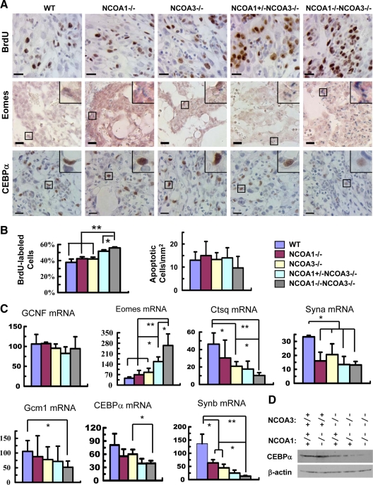
Figure 5.
Compound knockout of NCOA1 and NCOA3 increased proliferation and decreased differentiation of the labyrinth trophoblasts at E12.5. A, Detection of the BrdU-labeled proliferative cells (brown) by IHC (upper panels), the stem-like trophoblasts by in situ hybridization of Eomes mRNA (blue) (middle panels), and the CEBPα protein (brown) in layer III trophoblasts by IHC (lower panels) in the labyrinth layer of E12.5 placentas with the indicated genotypes. The insets at upper right corners are amplified images of the boxed areas. Scale bars, 20 μm. B, The percentage of BrdU-labeled labyrinth trophoblasts in total labyrinth trophoblasts (left panel) and the number of apoptotic cells per mm2 area of labyrinth with indicated genotypes. Apoptotic cells were detected by TUNEL assay. The cell numbers of three samples for each group were counted by using the ImageTool software. C, Relative mRNA levels of several labyrinth trophoblast marker genes assayed by qPCR. *, P < 0.05; and **, P < 0.01, compared by t test between any two groups (n = 3 for each group). D, Western blot analysis of CEBPα in E12.5 placentas of mouse embryos with the indicated genotypes. β-Actin served as a loading control. GCNF, Germ cell nuclear factor.

