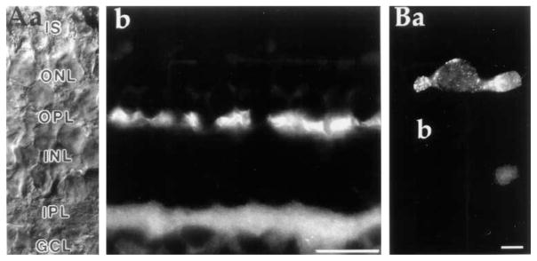
Figure 3. Antibodies to the Plasma Membrane Ca2+-ATPase Are Localized to the Inner Segments and Synaptic Terminals of Photoreceptors.
(A) Immunostaining of radially cut section of tiger salamander retina. Nomarski DIC image (a) and anti-PMCA immunofluorescence (b) of retinal section are shown. The strongest labeling occurred at regions of synaptic contacts in the outer and inner plexiform layers. Photoreceptor inner segments were also stained, albeit not as strongly. Scale bar, 40 μm.
(B) Example of an enzymatically dissociated cone labeled with a secondary antibody in the presence (a) and absence (b) of the primary PMCA antibody. The cells in (a) and (b) were plated and stained in parallel on concanavalin A–coated dishes. Scale bar, 10 μm.

