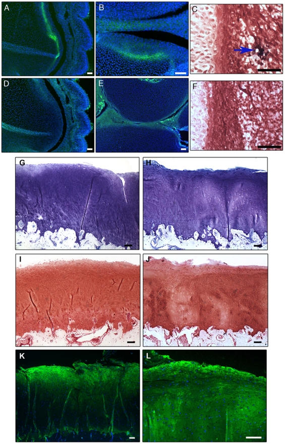
Figure 7. Engraftment of human cartilage progenitor cells into developing chick hind limbs.
Cells expressing human collagen type I are present in the growth plate of the developing chick limb at st36 (A). The high power image demonstrates human collagen type I expression in the surface region of the developing cartilage anlagen (B). IgG negative control (D). Expression of chick collagen type I is not evident in st36 chick limb (E). ISH for human Alu repeats on a representative section from a st36 chick limb (C). The Alu-positive nuclei are stained black (arrowed). Negative control for ISH for the Alu repeats (F). Scale bars = 50 µm. In vivo implantation of goat chondroprogenitors into cartilage defects. Histological stained sections of repair tissue in the caprine in vivo repair model. Toluidine blue stained sections of repair tissue containing the membrane seeded with full-depth chondrocytes (G) and chondroprogenitors (H) shows examples of an integrated repair tissue. Safranin O staining demonstrated proteoglycan synthesis in the repair tissue in defects treated with full-depth chondrocyte seeded membranes (I) and chondroprogenitor seeded membranes (J). Repair tissue in defects treated with chondrocyte seeded membranes (K) or chondroprogenitor seeded membranes (L), labelled positive for collagen type II. Scale bars = 100 µm.

