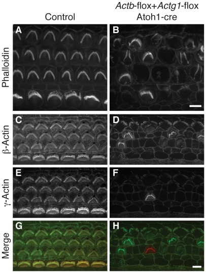
Figure 3. Actb Actg1 double knockout cells do not develop stereocilia.
(A, B) Phalloidin staining of organ of Corti from control (A) or Actb-flox Actg1-flox Atoh1-cre (double knockout) (B) P5 pups shows normal stereocilia in control but generally absent stereocilia in the double knockout. (C–H) double label with dye-conjugated antibodies to β-actin or γ-actin show that remaining stereocilia contain one of the actin isoforms. In merged images in (G–H) β-actin is green and γ-actin is red. Bars, 5 µm.

