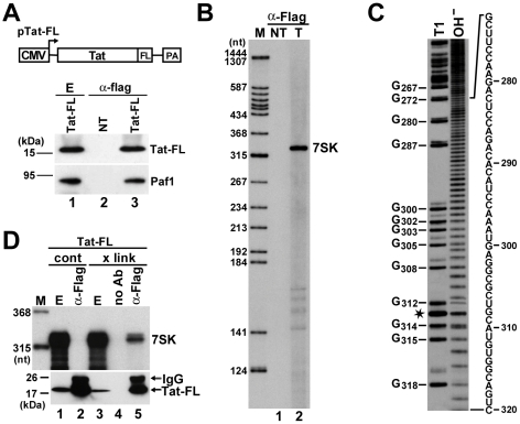
Figure 1. In vivo association of HIV Tat with 7SK.
A. Transient expression of Tat-FL in HeLa cells. Schematic structure of the pTat-FL expression construct is shown. The cytomegalovirus promoter (CMV) and the polyadenylation region (PA) are indicated. Tat-FL was immunoprecipitated (α-Flag) from extracts (E) prepared from transfected or non-transfected (NT) cells. Distribution of Tat-FL and Paf1 was monitored by Western blot analysis. B. Detection of Tat-associated HeLa RNAs. RNAs co-precipitated with Tat-FL (T) were labeled in vitro and separated on a 6% sequencing gel. Lanes NT and M, control IP from non-transfected cells and molecular size markers. C. RNA G-tracking. The Tat-associated RNA was partially digested with RNase T1 or moderately hydrolyzed with formamide (OH−) and analyzed on a 6% gel. G residues and their positions in the human 7SK snRNA sequence are indicated. Asterisk indicates a fragile U residue. D. In vivo cross-linking of Tat-FL and 7SK. HeLa cells expressing Tat-FL were treated (x link) or not treated (cont) with formaldehyde before extract (E) preparation. Tat-FL was immunoprecipitated (α-Flag) or mock-precipitated (no Ab) under stringent conditions. Distributions of Tat-FL and 7SK snRNA were monitored by Western blot analysis and RNase A/T1 mapping, respectively.

