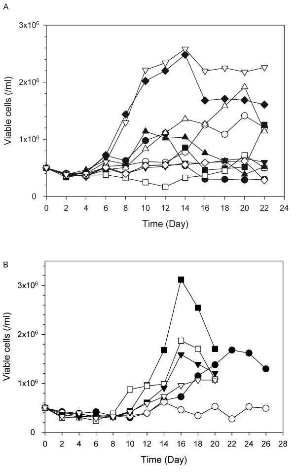
Figure 4.
Growth profiles of cells cultured in an optimized SFM. A. Comparison with SFM* shown in Figure 3A. Black circles, SFM* #1; white circles, SFM* #2; inverted black triangles, SFM* #3; white triangles, SFM* #4; black squares, SFM* #5; white squares, SFM* #6; black diamonds, SFM* #7; white diamonds, SFM* #8; black triangles, SFM* #9; inverted white triangles, optimized SFM. This figure shows one representative growth profile among the three independent cultures. B. Comparison with SCM. Three sets of cultures were performed independently with PBMCs obtained from the three different donors. Symbol indicates each set. Black symbols represent cells grown in optimized SFM, and white symbols represent cells grown in SCM.

