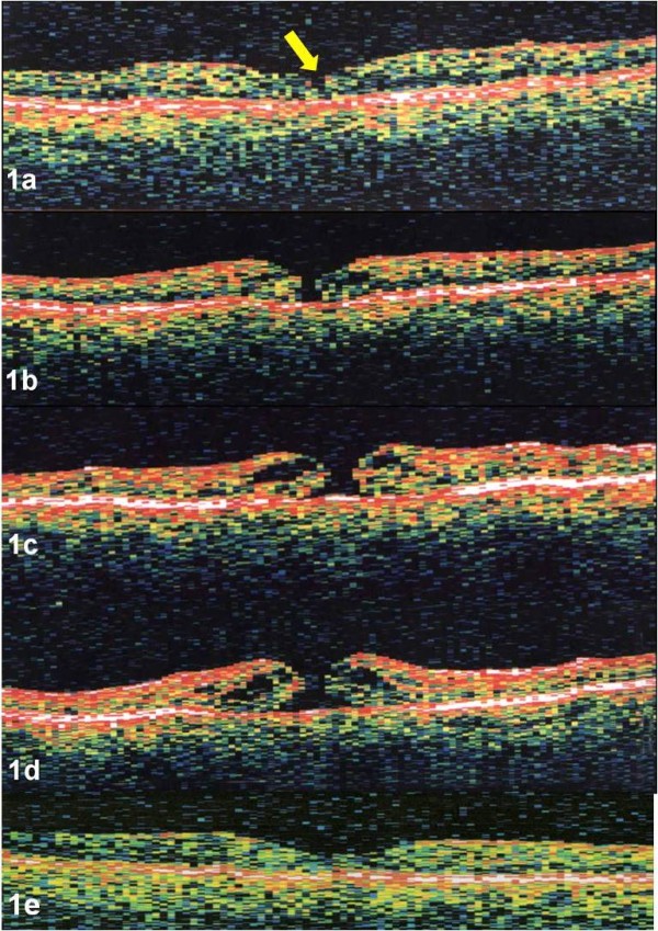
Figure 1.

a-e. Case 1. OCT image of the right eye demonstrated vitreofoveal separation with a small defect in the ILM (yellow arrow) in the center of the fovea (Figure 1a). Serial OCTs, performed ten days (Figure 1b), 3 weeks (Figure 1c), and six weeks (Figure 1d) later demonstrated progression to a stage 2 macular hole without evidence of traction. Post-operative OCT revealed closure of the macular hole with restoration of normal foveal contour (Figure 1e). OCTs were performed using OCT 2000, (Humphrey Instruments, San Leandro, CA)
