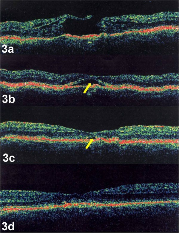
Figure 3.

a-d. Case 3. OCT image of the right eye demonstrated a full thickness stage 2 macular hole with underlying drusen (Figure 3a). Post-operative OCT images, performed 2 weeks (Figure 3b), 4 weeks (Figure 3c), and 8 weeks (Figure 3d) after surgery, demonstrated closure of the hole with absorption of the sub-retinal fluid (yellow arrow). OCTs were performed using OCT 3000, (Humphrey Instruments, San Leandro, CA)
