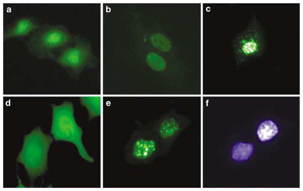
Figure 1.
Cellular localization and toxicity of GFP-apoptin in primary endothelial and MCF-7 cells. HUVECs (a–c) and MCF-7 cells (d–f) were transiently transfected with the pEGFP-C1 control vector (a, d) or with a pEGFP-apoptin construct (b, c, e, f). The pictures were taken 24 h (a, b, d, e, f) and 96 h (c) post-transfection. (f) DAPI staining of cells shown in (e).

