
Figure 2.
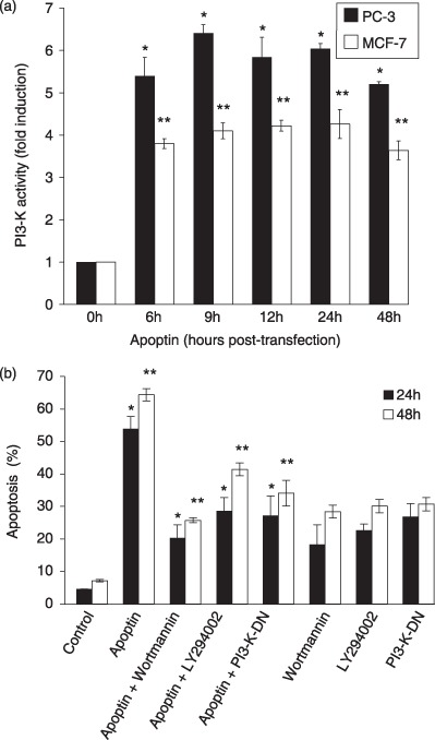
PI3‐K is constitutively activated during apoptin‐induced apoptosis. (a) PI3‐K activity was measured by an ELISA‐based assay after immuno‐precipitating PI3‐K from the lysates of PC‐3 and MCF‐7 cells, transfected to express apoptin (time points indicate time post‐transfection) as described in the methods section, and the fold induction was calculated. PI3‐K activity in non‐transfected cells was considered as a basal level (1x). Significance of the data was statistically confirmed (*P < 0.02 and **P < 0.04) using Student's t‐test. (b) The effect of PI3‐K inhibition on apoptin‐induced cell death was assessed by flow cytometry (Nicoletti method). Cells were either transfected to express GFP (control) or GFP‐apoptin, in the absence or presence of treatment with wortmannin or LY294002, or transfection to co‐express the dominant‐negative PI3‐K (PI3‐K‐DN). Apoptosis was then measured 24 and 48 h post‐transfection, with results compared to those for control treatments of wortmannin or LY294002 alone, or PI3‐K‐DN expression alone. The data are statistically significant (*P < 0.02 and **P < 0.03) as revealed by Student's t‐test.