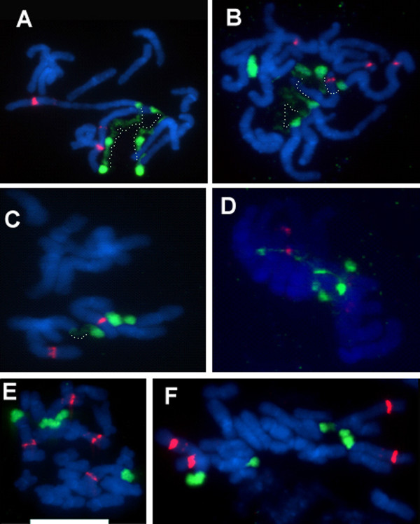
Figure 4.
FISH to metaphase spreads of parental diploid species (A-D) and synthetic T. miscellus lines (E-F). (A) T. pratensis 2608. (B) T. porrifolius 2607. (C) T. dubius 2613. (D) T. porrifolius 2611. (E) and (F) stand for the 111-1 and 111-4 synthetic individuals, respectively. Metaphases were hybridized with the 18S rDNA (painting 35S sites in green) and 5S rDNA (red fluorescence) probes. Note the discontinuous chromatin condensation along the loci. Regions of condensed and decondensed chromatin are interconnected with dotted lines in (A-C). Scale bar = 10 μm.

