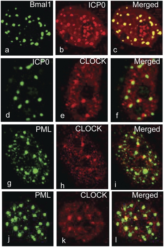
Fig. 1.
In infected cells CLOCK and Bmal1 localize at ND10 bodies. (A–C) HEp-2 cells were exposed to 10 pfu of HSV-1(F) per cell 24 h after transfection of a plasmid encoding HA–Bmal1. The cells were fixed 6 h after infection and reacted with the mouse monoclonal antibody to HA and the rabbit polyclonal antibody to ICP0. (D–F) HEp-2 cells were transfected with the empty vector pcDNA3.1Zeo(+) for 24 h prior to infection with the HSV-1(F) (10 pfu/cell). The cells were fixed at 6 h after infection and the cells reacted with the rabbit polyclonal antibody against CLOCK and the mouse monoclonal antibody against ICP0. (G–I) HEp-2 cells were infected with the wild-type virus (10 pfu/cell). The cells were fixed at 4 h after infection and reacted with the rabbit polyclonal antibody against CLOCK and the mouse monoclonal antibody against PML. (J–L) Mock-infected HEp-2 cells fixed and reacted with antibody as above. All of the images were captured with the same settings of a Zeiss confocal microscope.

