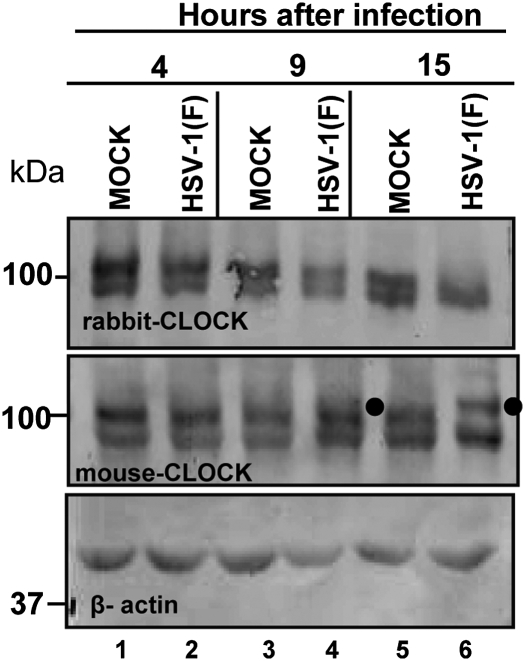
Fig. 2.
Clock is modified in HSV-1(F)–infected cells. HEp-2 cells were mock infected or exposed to 10 pfu of HSV-1(F) per cell. The cells were harvested and lysed at 4, 9, or 15 h after infection. The electrophoretically separated denatured proteins were transferred to nitrocellulose sheets and immunoblotted either with the rabbit CLOCK antibody (Upper) or with the mouse CLOCK antibody (Lower). β-Actin served as a loading control. The filled circles point to a modified form of CLOCK.

