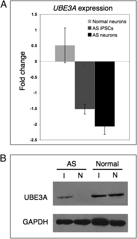
Fig. 4.
Paternal UBE3A is repressed in AS iPSC-derived neurons. (A) qRT-PCR analysis of UBE3A expression in AS and normal iPSCs and iPSC-derived neurons. Gene expression is normalized to GAPDH and is presented as the fold change relative to UBE3A expression levels in normal iPSCs. Error bars indicate SD for three independent cultures. (B) Western blot analysis of normal and AS iPSCs (I) and 10-wk-old iPSC-derived neurons (N).

