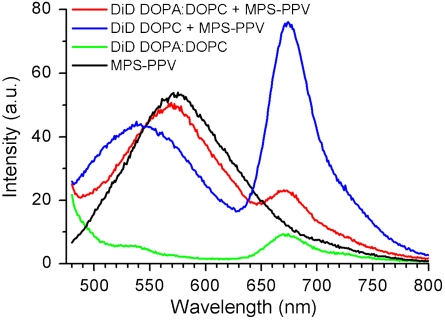
Fig. 2.
Emission spectra upon 457-nm excitation of 1.6 × 10⁻⁵ M MPS-PPV in monomer units with (red) DOPC vesicles stained with DiD (250∶1 lipid∶DiD ratio, solutions were 0.20 mM in lipid); (black) DOPA∶DOPC (3∶1) vesicles stained with DiD (250∶1 lipid∶DiD ratio, solutions were 0.20 mM in lipid). Also shown is the emission of free MPS-PPV (blue) and of DOPA∶DOPC (3∶1) vesicles stained with DiD (250∶1 lipid∶DiD ratio, solutions were 0.20 mM in lipid) (green).

