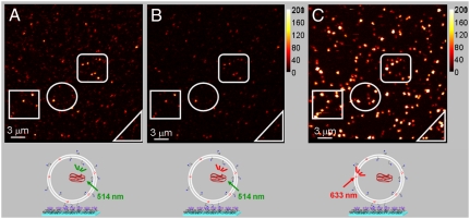
Fig. 4.
Fluorescence scanning confocal images for MPS-PPV encapsulated in anionic vesicles (DOPA∶DOPC; 3∶1) containing ca. 20 DiD/vesicle. The vesicles were prepared as described in the caption of Fig. 3 and further separated from free polymer via gel filtration chromatography and diluted to a final 100-pM concentration prior to surface immobilization. A (green channel) and B (red channel) were acquired simultaneously upon 514-nm excitation of MPS-PPV. C was obtained upon directly exciting vesicle embedded DiD with the 633-nm line of a HeNe laser. The right bar illustrates the counts per millisecond per pixel.

