Fig. 1.
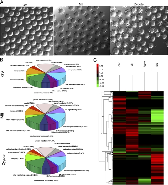
Total proteins identified in mouse oocytes and zygotes. (A) Representative images of mouse oocytes at different developmental stages including GV, MII, and zygote. As shown, fully grown GV oocytes with visible germinal vesicles, MII oocytes with the first polar bodies, and zygotes with two pronuclei were carefully selected. (B) Functional categorization of the identified proteins in GV and MII oocytes and zygotes. The identified proteins were grouped into 14 categories according to their molecular functions, which include involvement in the cell cycle and proliferation, transport, stress response, developmental processes, RNA metabolism, DNA metabolism, cell organization and biogenesis, cell–cell signaling, signal transduction, cell adhesion, protein metabolism, death, other metabolic processes, and other biological processes. We then calculated the percentage of every group of proteins. (C) Clustering of proteins in GV and MII oocytes, zygotes, and ES cells with a color gradient for gene abundance ranks. The cluster of more abundantly expressed proteins is highlighted in red. The total number of observed spectra assigned to each protein in each cell type was used as the basis for clustering. Clustering showed that the protein expression profile is similar for MII oocytes and GV oocytes, whereas the profile of zygotes is more similar to ES cells.

