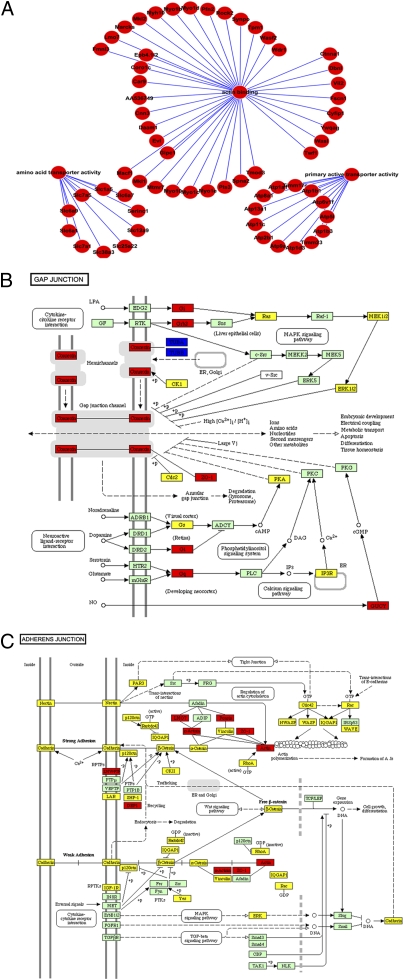
Fig. 2.
Highly expressed proteins in GV oocytes. (A) Actin binding proteins (shown at the top), primary transporters (shown at the left), and amino acid transporters (shown at the right) are more abundantly expressed in GV oocytes compared with MII oocytes and the zygotes. (B) The gap junction pathway is overrepresented in the GV oocyte. Red rectangles represent the gap junction proteins expressed more abundantly in GV oocytes compared with MII oocytes. Yellow rectangles represent the comparable expression level of gap junction proteins between GV and MII oocytes. Green is the color of Kyoto Encyclopedia of Genes and Genomes (KEGG) database. (C) The adhesion junction pathway is overrepresented in the GV oocyte. Red rectangles represent the adhesion junction proteins expressed more abundantly in GV oocytes compared with MII oocytes. Yellow rectangles represent the comparable expression level of adhesion junction proteins between GV and MII oocytes. Green is the color of KEGG database.

