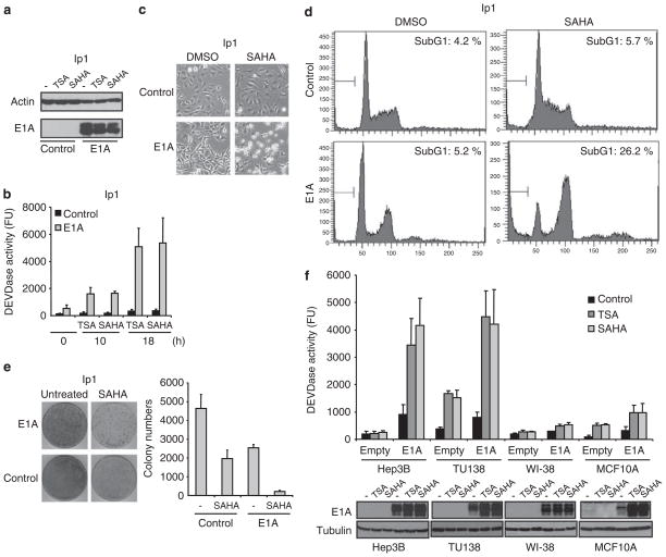
Figure 1.
Adenovirus 5 early region 1A (E1A) enhances HDACi-induced cell death in human cancer cells. (a) E1A and actin expression in SKOV3-ip1 control or E1A cells was verified by immunoblot analysis. (b) SKOV3-ip1 control or E1A cells were treated with 250 nM of trichostatin A (TSA) or 5 μM of suberoylanilide hydroxamic acid (SAHA) for the indicated periods, and caspase-3 activity was determined by caspase assay, using a fluorescence substrate. (c) The cellular morphology of SKOV3-ip1 control or E1A cells treated with dimethyl sulfoxide (DMSO) or 5 μM of SAHA for 24 h. (d) SKOV3-ip1 control or E1A cells were treated with DMSO or 5 μM of SAHA for 20 h. The cells were then fixed and stained with propidium iodide, followed by flow cytometric analysis. The percentage of cells with a sub-G1 DNA content is shown within each box. (e) SKOV3-ip1 control or E1A cells were treated with 5 μM of SAHA for 24 h. Cellular sensitivity to SAHA was determined by using the clonogenic survival assay. The colony numbers were counted and shown in the bar graph (n=3). (f) Hep3B, TU138, WI-38 and MCF10A cells were transiently transfected with either empty or E1A expression plasmid and treated with 250 nM of TSA or 5 μM of SAHA for 16 h. Capase-3 activity was then determined by caspase assay using fluorescence substrate. E1A and tubulin expression in these cells was verified by immunoblot analysis.

