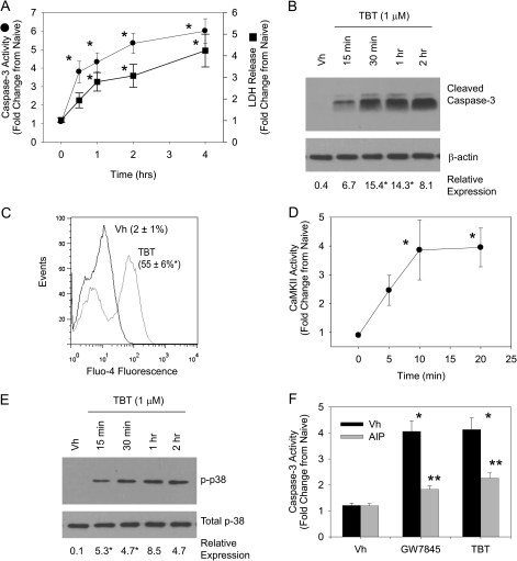
FIG. 6.
The immunotoxicant TBT induces calcium-dependent cell death. BU-11 cells were treated with Vh (DMSO, 0.1%) or TBT (1μM) for 5 min 2 h. (A) Caspase-3 activity and LDH release were assessed using the Caspase3/7-Glo Assay and the CytoTox-Glo Assay (Promega). Luminescence values in experimental wells were normalized by that measured in untreated cells and are presented as “Fold Change from Naïve.” (B) Formation of cleaved active caspase-3 (17 kDa) was analyzed by immunoblotting of cytoplasmic extracts. Protein expression was quantified as described in the Materials and Methods and presented as “Relative Expression.” (C) Cytoplasmic Ca2+ was detected by loading BU-11 cells with Fluo4-AM (1μM) prior to treatment with Vh or TBT for 15 min, followed by flow cytometry. (D) CaMKII activity was determined in cytoplasmic extracts using the SignaTect Assay (Promega). CPM values in experimental wells were normalized by that measured in untreated cells and are presented as “Fold Change from Naïve.” (E) p38 MAPK activation was determined by detection of phosphorylated p38 MAPK in cytoplasmic extracts by immunoblotting. (F) BU-11 cells were pretreated with Vh (DMSO, 0.1%) or AIP-II (4μM) for 30 min and then treated with Vh (DMSO, 0.1%) or TBT (1 μM). Caspase-3 activity was assessed using the Caspase3/7-Glo Assay, as described in (A). Data are presented as means ± SE from at least three independent experiments. *Statistically different from Vh-treated (p < 0.05, ANOVA, Tukey-Kramer). ** Statistically different from GW-treated alone (p < 0.05, ANOVA, Tukey-Kramer).

