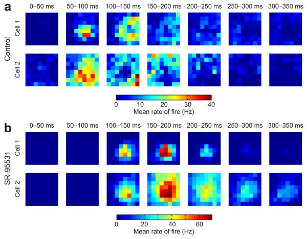
Figure 7.
GABAergic circuits reduce spatiotemporal correlations in tectal receptive fields. (a) Receptive fields were analyzed in the temporal domain by separating responses according to when they occurred after the onset of the flashed stimuli. Data from two representative control cells are shown for seven different time bins (each 50 ms long) following the onset of the stimuli. As a population, control cells showed greater variety in their responses at different times and different locations of visual space (n = 21 cells). Associated with this variability was the fact that stimuli in some regions of a control cell's receptive field would typically elicit early responses (for example, 50–100 ms post-stimulus), whereas stimuli in other locations would generate later responses (for example, 200–250 ms post-stimulus). (b) In contrast with control cells, the population of cells recorded under GABAA receptor blockade showed similar responses at different times and locations of visual space (n = 7 cells). This is illustrated with data from two representative SR-95531 cells. Maximal responses for the SR-95531 cells occurred between 100–200 ms in post-stimulus time and typically the same receptive field locations elicited both early spikes and later spikes. The result was greater uniformity in the spatiotemporal profile of the responses of different cells in the SR-95531 condition.

