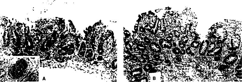
Figure 11.
Acute vascular rejection of positive cross-match isolated intestinal graft. (A) This biopsy specimen obtained 4 hours after transplantation shows typical changes of acute vascular rejection, with pronounced hemorrhage and congestion obscuring the lamina propria. The villi, although intact, are shortened, and the surface epithelium is focally lost. Inflammatory infiltration is minimal. Small arterioles demonstrated endothelial hypertrophy, mural thickening, platelet thrombi, and focally sparse neutrophil infiltration (insert). (B) Fifteen days after transplantation and after treatment with steroids and OKT3, the mucosa displays prominent evidence of mucosal regeneration. The crypts are irregular, branched, and lined by hyperplastic epithelium with crowded, pseudostratified cells. Toward the luminal surface, early villus formation can be appreciated. The lamina propria retains a mild component of inflammatory cells, primarily small lymphocytes and scattered plasma cells.

