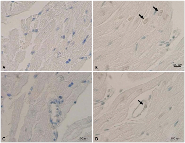
Fig. 3.
In situ labeling of DNA fragmentation in rat myocardium from TUNEL assay. Fragmented DNA is labeled in brown, and all nuclei are counterstained with methyl-green. Myocardial sections from rats in the control group reveal no evidence of DNA fragmentation in cardiomyocytes (A) or endothelial cells (C). A heterogenous pattern is revealed for the isolated apoptotic nuclei in the myocardium of doxorubicin-treated rats, as indicated by arrows, for both cardiomyocytes (B) and endothelial cells (D). TUNEL: terminal deoxynucleotidyl transferase-mediated dUTP nick-end labeling.

