Figure 10.

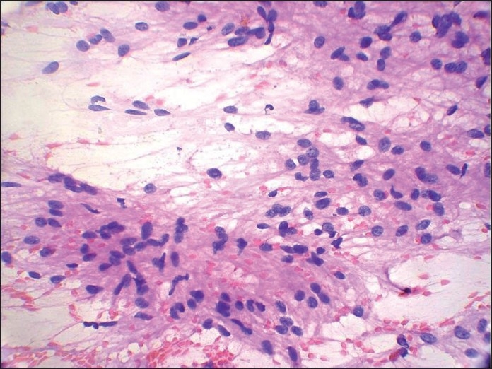
Papanicolaou-stained smear shows spindle cells showing reduced intercellular cohesion with individual cells escaping from the surface of fragments suggesting a malignant lesion. Palisading is seen at places

Papanicolaou-stained smear shows spindle cells showing reduced intercellular cohesion with individual cells escaping from the surface of fragments suggesting a malignant lesion. Palisading is seen at places