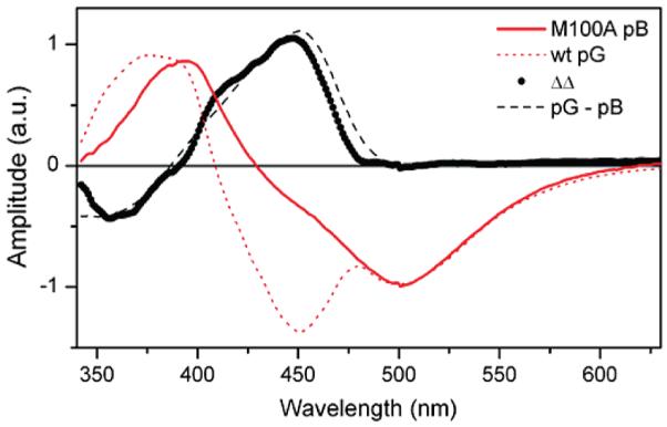
Figure 3.

Comparison of 15 ps EADS from the pB state of M100A (red, solid) with SADS obtained from photoexcitation of the pG state in wild-type PYP (red, dotted). The difference spectrum ΔΔ (black, circles) indicates that the pB and pG states have identical picosecond emission spectra, suggesting that the predominate fluorescent excited-state species in photoexcited pB has a deprotonated chromophore. It resembles the static difference spectrum of pG and pB (black, dashed).
