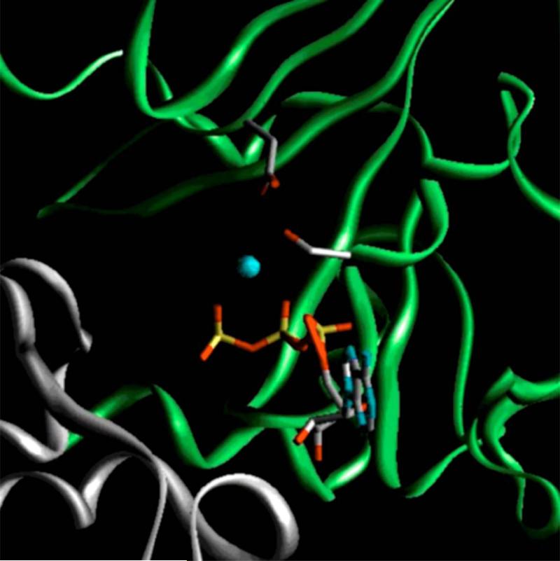
Fig. 1. Model of Mg·ATP-bound form of wild-type nucleotide binding site 1 of human MRP1.
The nucleotide binding domain 1 (NBD1, 643-855) of human MRP1 was modeled based on the crystal structure of Mg·ATP-bound form of wild-type MRP1-NBD1 [30], whereas the NBD2 was modeled using the MJ0796-E171Q homodimer [17] as a template. The gray ribbon indicates the residues from NBD1, whereas the green ribbon represents the residues from NBD2. The gray sticks on the gray ribbon represent S685 in Walker A motif and D792 in Walker B motif. ATP is shown in stick representation. Red indicates oxygen atom; blue, nitrogen; yellow, phosphorus; cyan, Mg++ ion.

