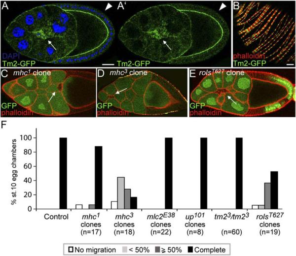
Figure 5. Muscle-Specific Genes and Border Cell Migration.
(A and B) (A) Tropomyosin2-GFP (protein trap) in migrating border cells (arrow) compared to follicle cells (arrowhead) and (B) in the muscle sheath that surrounds egg chambers, counterstained for F-actin (phalloidin, red). The scale bars are 20 μm.
(C) Stage-10 egg chamber with an mhc1 mutant border cell clone (marked by the absence of GFP) that shows normal migration.
(D) Stage-10 egg chamber with an mhc3 mutant border cell clone (marked by the absence of GFP) that is delayed (<50%).
(E) Late stage-9 egg chamber with a rolsT627 mutant border cell clone that shows defective migration (50%).
(F) Quantification of border cell migration in egg chambers at stage 10 for mhc1, mhc3, mlc2E38, up101 and rolsT627 mutant border cell clones, or in tm23 homozygous mutant females. Controls for mhc1, mhc3, and rolsT627 mutant clones were: mhc1/+ (n = 46), mhc3/+ (n = 21), and rolsT627/+ (n = 20).

