
Figure 1.
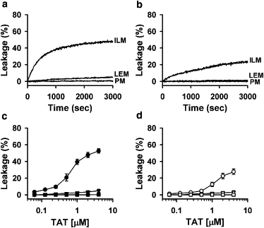
TAT peptide releases the encapsulated probe from ILM and LEM liposomes, but not from CPM liposomes. (a and b) Kinetics of dye dequenching due to the release of ANTS/DPX from 25 μM liposomes of ILM, LEM, and PM lipid composition upon addition of 2 μM TAT peptide at pH 5.5 (a) and pH 7.4 (b). (c and d) Dependence of dye dequenching, measured 50 min after the addition of TAT, on peptide concentration. Peptide was added to 25 μM of ILM (circles), LEM (triangles), and PM (squares) liposomes at pH 5.5 (c) and at pH 7.4 (d). Each data point in panels c and d represents the mean of four independent experiments, and error bars indicate the standard deviation (SD).