
Figure 2.
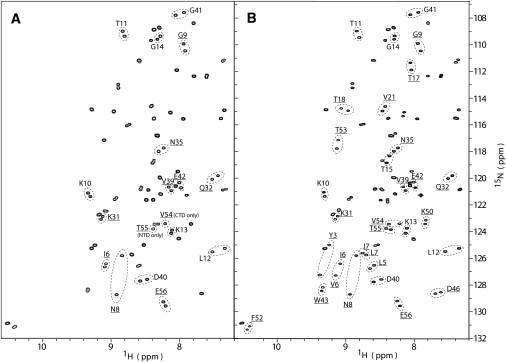
1H-15N HSQC spectra of dGB1 domain proteins. (A) 1H-15N HSQC spectrum of dGB1L7I-(6)-GB1. Pairs of resonances that are distinguishable between the GB1L7I (NTD) and the GB1 (CTD) are labeled with the residue name and number and circled. Ten pairs with 15N-{1H} NOE values > 0.65 were used for diffusion tensor determination (underlined). These pairs are from identical residues residing on the NTD and CTD, respectively, with the exception of T55 (NTD) and V54 (CTD), which possess no resolved counterpart in the other domain. The spectrum of the dGBL7I-(6)-GB1 protein is almost indistinguishable from those of the single-domain proteins sGB1 and sGB1L7I. (B) 1H-15N HSQC spectrum of dGB3β1-(6)-GB1. Thirty labeled pairs of resolved resonances are present, and 22 of these pairs exhibit 15N-{1H} NOE values > 0.65.