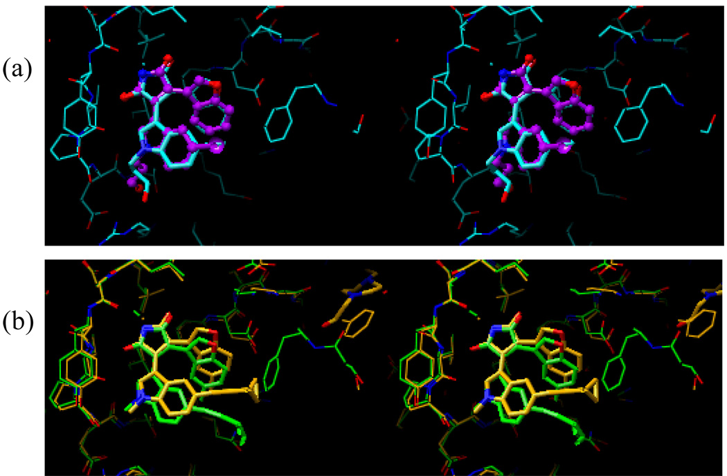
Figure 10.
Superposition of the docked and the preliminary X-ray crystal structures of compounds 5 (a) and 14 (b). In Figure 10a, the docked structure of compound 5 is shown in purple, while the X-ray crystal structure of the same compound is shown in cyan. In Figure 10b, the docked structure of compound 14 is shown in green, while the X-ray crystal structure of the same compound is shown in orange.

