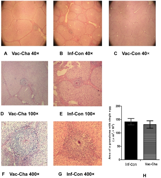
Figure 3. Liver histology at 8 weeks after infection with HE staining and the average area of a single granuloma containing one egg.
Figures 3A, 3D and 3F show fewer granulomas and mild inflammation in the livers of the Vac-Cha group. Figures 3B, 3E and 3G show severe granulomatous inflammation in the livers of the Inf-Con group. Figure 3C shows no granuloma in the livers of the Vac-Con group. Figure 3H shows the average area of a single liver granuloma containing only one egg in the Vac-Cha and Inf-Con groups (n = 7).

