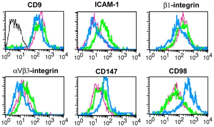
Figure 1. Flow cytometry analysis of the expression of cell adhesion molecules in endometrial epithelial cells and cell lines.
Histogram profiles of the plasma membrane expression of CD9 (VJ1/20), ICAM-1 (HU5/3), β1 integrin (TS2/16), αvβ3 integrin (8D6), CD147 (VJ1/9) and CD98 (FG1/10) on HEC-1-A (green) and RL95-2 (blue) cell lines as well as on primary EEC cultures (pink). Negative control pX63 is shown in gray line.

