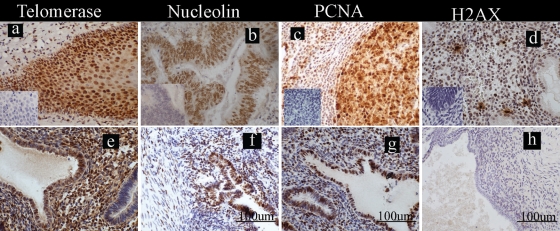
Figure 1.
Localization of the markers of cell fate studied in the ectopic endometrial lesions from women. Photomicrographs are representatives of the immunostaining for telomerase, nucleolin, proliferating cell nuclear antigen (PCNA) and histone γ-H2AX; positive staining = brown; scale bars = 100 µm (f, g and h) applicable to all panels. (a–d) External positive control tissues with inserts illustrating the immunologically negative controls: (a) telomerase in human tonsillar cortex, (b) nucleolin in human adeno-carcinoma of the colon, (c) PCNA in human tonsil and (d) histone γ-H2AX in human late-secretory endometrium; (e–h) ectopic blue/chocolate lesions from women with endometriosis stained with (e) telomerase (f) nucleolin, (g)PCNA and (h) γ-H2AX.

