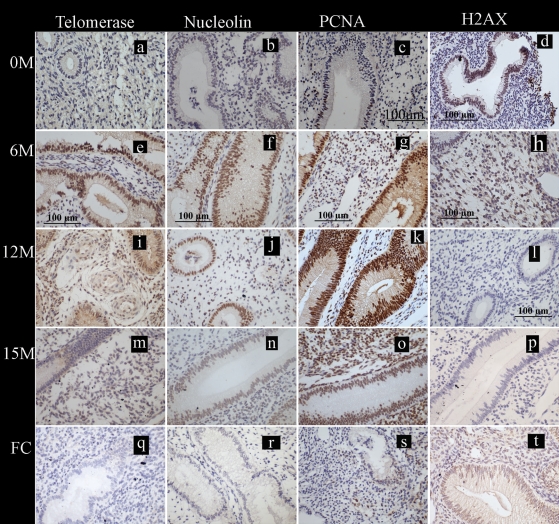
Figure 4.
Localization of the markers of cell fate studied in the eutopic endometrium collected during the WOI in baboons. Photomicrographs are representatives of the immunostaining for telomerase, nucleolin, proliferating cell nuclear antigen (PCNA) and histone γ-H2AX; positive staining = brown; scale bars = 100 µm (c–h and l) applicable to all panels. (a–d) Endometrium from baboons (n = 5) prior to inoculation at 0 M and stained for telomerase, nucleolin, PCNA and histone γ-H2AX. (e–p) Eutopic endometrium from animals with induced disease (6, 12 or 15 months post-induction tissue from six animals included at each time point) stained with telomerase, nucleolin, PCNA and γ-H2AX. (q–t) Eutopic endometrium from the control healthy fertile animals (n = 8) stained for telomerase, nucleolin, PCNA and γ-H2AX.

