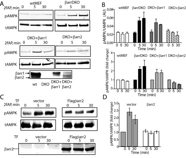
Figure 4.
Expression of β-arrestin-2 inhibits PAR2 stimulated AMPK activation. A, B. Mouse embryonic fibroblasts from wt embryos (wtMEF), β-arrestin double knockout embryos (MEF βarrDKO), or MEFβarrDKO stably expressing βarrestin-1 (DKO+βarr1) or β-arrestin-2 (DKO+βarr2) were treated with 2fAP for 0, 5 or30 minutes and analyzed as described in Figure 1. A. Representative western blots of pAMPK and tAMPK in each cell line are shown in the upper two panels. The lower panel shows β-arrestin expression in each cell line. B. Bar graphs depicting normalized phospho-AMPK levels (upper) and fold changes over baseline in pAMPK/tAMPK (lower) in each cell line. Baseline is defined as pAMK observed in the absence of 2fAP for each cell line. C. Cells were transfected with an empty vector or flag-β-arrestin-2, treated with 2fAP for 0, 5 or 30 minutes and analyzed as described above. Upper panels: anti-pAMPK and tAMPK; lower panels: anti-β-arrestin was used to confirm expression of transgene. D. Bar graph showing fold changes compared with baseline in pAMPK in cells transfected with vector or Flagβarr2. Baseline in these experiments is defined as pAMPK in untreated, vector transfected cells.

