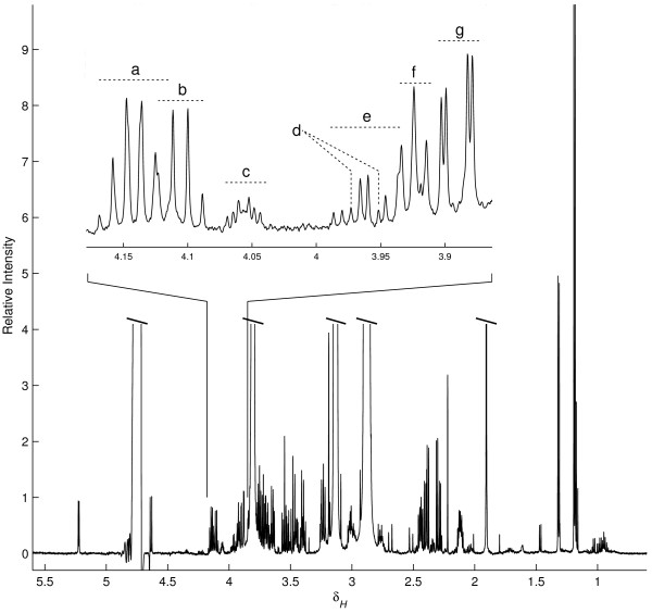
Figure 5.
Typical 600 MHz 1H NMR spectrum (δ 0.5-5.6) of a male rat liver perfusate sample. Resonances from the buffer, HEPES and acetate, as well as the residual water signal were cropped to reveal the more interesting liver-derived signals. The middle expansion shows in detail a small portion of the ppm-axis. Assigned signals in the expanded region are (a) 3-hydroxybutarate, (b) lactate, (c) choline, (d) histidine, (e) serine, (f) HEPES (13C-satelite), and (g) glucose.

