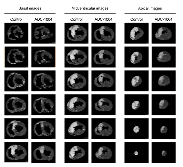
Figure 2.
Infarct size. Delayed contrast enhanced MR images from one typical animal from each group. Approximately 200 short-axis images, each 0.5 mm thick, are analyzed from every heart. Infarcted myocardium (white) is defined as hyper-enhanced myocardium with signal intensity above eight standard deviations of the signal intensity in the remote myocardium. Microvascular obstruction is defined as hypointense regions in the core of the infarction with signal intensity less than the threshold for infarction.

