
Figure 6. The glutamate receptor GLR-1 accumulates in cand-1 mutants.
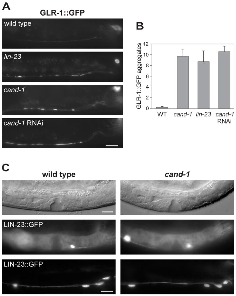
(A) Epifluorescence images of GLR-1∷GFP signal in the posterior ventral nerve cord of wild-type, lin-23(e1883) mutant, or cand-1 (tm1683) mutant young adults. Note that the signal from nerve cell bodies (upper right) is out of focus in several of the images. (B) A graph of the number of GLR-1∷GFP aggregates per ventral nerve cord that are ≥ 2 μm in length, n=10 animals. (C) Analysis of LIN-23∷GFP levels in wild type and cand-1(tm1683) mutants. The vulva and gonadal regions of early L4-stage stage animals are shown as DIC images (top) or LIN-23∷GFP epifluorescence images (middle). LIN-23∷GFP epifluorescence in the posterior ventral nerve cord of young adults are shown in the bottom panels. Posterior is to the right in all images. Scale bars, 10 μm.