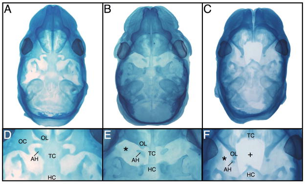
Figure 2.
Whole mount stained Theiler Stage 24 (E16) crania (A–F). The +/+ head (A,D) displays normal cranial base development while the Br/+ (B,E) cranium shows a fused trabecular cartilage lacking lateral chondrification, and the orbital cartilages are absent (*). Although the hypochiasmatic cartilages are present, they contact the orbitonasal laminae abnormally. The Br/Br cranium (C,F) displays unfused trabecular cartilages that are truncated caudally so that they do not meet the hypohyseal cartilage forming an abnormal rostral point. The orbitonasal laminae are displaced laterally and abnormally abut the reduced hypochiasmatic cartilages while the orbital cartilages are absent. AH = hypochiasmatic cartilage; HC = hypophyseal cartilage; OC = orbital cartilage; OL = orbitonasal lamina; TC = trabecular cartilage.

